What is Folliculometry?
Ovulation is a key part of a woman’s menstrual cycle, where an egg is released from the ovary. Problems with ovulation are a common cause of infertility in women. In 1978, Dr. Joachim Hackelöer introduced the use of ultrasound to observe the growth of follicles—small sacs in the ovaries that contain eggs—during the menstrual cycle.
Later, a technique called transvaginal sonography (TVS) was developed. In this method, a small ultrasound probe is inserted into the vagina, bringing it closer to the ovaries and uterus.
The benefits of transvaginal ultrasound include:
- Better Visualisation of Ovaries
- Early Detection of Follicles: It allows doctors to see and track small follicles as tiny as 2-3 millimetres throughout the menstrual cycle.
- No Need for a Full Bladder • Higher Image Quality
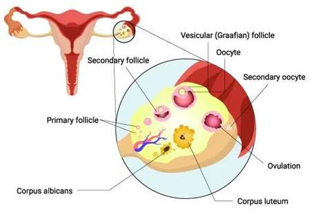
Folliculometry, also known as follicular monitoring, is a series of ultrasound scans used to track the development of ovarian follicles—small sacs in the ovaries that contain eggs—throughout a woman’s menstrual cycle. This process helps identify the best time for conception and is commonly used in natural cycles, as well as during fertility treatments like Intrauterine Insemination (IUI) and In Vitro Fertilisation (IVF).
Typically performed via transvaginal ultrasound, folliculometry usually begins on day 2 or 3 of the menstrual cycle and continues until ovulation is confirmed. The monitoring process is divided into three main phases:
- Baseline Scan: Conducted on day 2 or 3 of the cycle, this initial scan assesses the ovaries and uterus in their resting state. It provides important information such as the antral follicle count (indicating ovarian reserve), and checks for any ovarian cysts, fibroids, or other abnormalities.
- Preovulatory Scan: Starting around day 9 or 10, this series of scans monitors the growth of the developing follicles. A dominant follicle is expected to grow approximately 1-2 mm per day, reaching about 17-25 mm in diameter when it’s ready to ovulate. The endometrial lining is also evaluated to ensure it is thickening appropriately to support potential implantation.
- Luteal Phase Scan: After ovulation, this scan confirms that the dominant follicle has released its egg, indicated by the collapse of the follicle and the presence of fluid in the pelvic cavity. This phase helps verify that ovulation has occurred successfully.
Follicular phase assessment via ultrasound scan
Baseline Scan:
A baseline ultrasound scan is an important procedure performed at the start of a woman’s menstrual cycle, typically on day 2 or 3. This transvaginal ultrasound provides valuable insights into reproductive health and is especially useful for those considering fertility treatments like Intrauterine Insemination (IUI) or In Vitro Fertilization (IVF).
Purpose of the Baseline Scan:
What is Ovarian Reserve:
This refers to the number of eggs a woman has available. By counting the small antral follicles (each measuring between 2–10 mm) in the ovaries, doctors can estimate ovarian reserve. A higher count suggests a better response to fertility treatments.
What is Ovarian Response:
This indicates how the ovaries might react to fertility medications. Understanding this helps in tailoring the appropriate drug dosage for treatments like IUI or IVF.
Key Assessments During the Baseline Scan:
- Ovarian Size and Volume: The size of the ovaries can provide clues about their function. Ovarian volume is calculated using the formula: length × width × height × 0.523. Typically, a combined ovarian volume of about 10 ml is considered normal. Smaller ovaries, with a volume less than 4 ml, may indicate a reduced response to stimulation.
- Ovarian Position: The scan checks if the ovaries are in their usual location. Sometimes, conditions like endometriosis or pelvic inflammatory disease (PID) can cause the ovaries to be displaced or adhere to other structures.
- Detection of Cysts or Other Issues: The scan can identify various ovarian cysts (such as functional cysts, endometriotic cysts, or dermoid cysts) and assess the health of surrounding structures like the fallopian tubes.
- Antral Follicle Count (AFC): Counting the number of antral follicles helps estimate ovarian reserve. An AFC of fewer than 4 follicles may predict a poor response to stimulation, while more than 16 follicles suggest a higher response. Combining AFC with blood tests like Anti-Müllerian Hormone (AMH) and Follicle-Stimulating Hormone (FSH) offers a comprehensive view of ovarian health.
Advantages of AFC (Antral Follicle Count):
- Age Correlation: AFC declines with age, providing insight into reproductive lifespan. Studies indicate a steady decrease in AFC as women age. For instance, research has shown that the number of small antral follicles (2–6 mm) declines with age, suggesting that AFC is a reliable indicator of diminishing ovarian reserve over time.
- Cost-Effective Assessment: Performing an AFC via ultrasound is a relatively affordable method for evaluating ovarian reserve and predicting how the ovaries might respond to stimulation in fertility treatments. It provides immediate information without the need for more invasive or expensive procedures.
- Enhanced Prediction with Combined Measures: When AFC is combined with assessments of ovarian stromal blood flow using color Doppler ultrasound, it may improve the prediction of ovarian response during In Vitro Fertilization (IVF) cycles. Some studies suggest that evaluating blood flow within the ovarian tissue can offer additional insights into ovarian function.
Clinical Application:
In stimulated fertility cycles, such as those involving Assisted Reproductive Technology (ART), combining AFC with factors like age, ovarian volume, and other ultrasound parameters helps in customizing the dosage of gonadotropins. This tailored approach aims to optimize ovarian response and improve the chances of successful treatment outcomes.
Pre-Ovulatory Scan:
A pre-ovulatory scan involves a series of ultrasound examinations during the follicular phase of the menstrual cycle to monitor the growth of ovarian follicles and predict ovulation timing. The schedule for these scans varies depending on the type of cycle:
- Natural or Intrauterine Insemination (IUI) Cycles: Monitoring typically begins around day 9 or 10 of the menstrual cycle.
- IVF Cycles: Monitoring often starts earlier, around day 5 or 6, with subsequent scans to closely track follicular development.
Purposes of Serial Scans:
- Evaluate Ovarian Response to Stimulation: Assess how the ovaries react to fertility medications, ensuring that the response is appropriate for the treatment plan.
- Monitor Pituitary Down-Regulation: Check the effectiveness of medications aimed at suppressing the pituitary gland to control hormone release during ART cycles.
- Prevent Ovarian Hyperstimulation Syndrome (OHSS): Regular scans help detect excessive ovarian responses early, allowing adjustments to medication to reduce the risk of OHSS.
- Track Follicle Development:
- Identify Growing Follicles: Monitor follicles as they develop, noting characteristics such as:
- Diameter exceeding 10mm.
- Growth rate of 2-3mm per day.
- Absence of internal echoes on ultrasound.
- Thin, regular walls.
- Measure Mature Follicles: Assess follicles around days 13-14 in a regular cycle, looking for:
- Size of 16-18mm.
- Thin walls.
- Presence of a thin, hypo-echoic rim in 30-40% of cases.
- Occasional visibility of the cumulus structure, indicating readiness for ovulation.
- Identify Growing Follicles: Monitor follicles as they develop, noting characteristics such as:
- Determine Timing for hCG Administration: By observing follicle maturity and optimal features, clinicians can accurately time the human chorionic gonadotropin (hCG) injection to trigger ovulation, crucial for procedures like IUI or egg retrieval in IVF.
- Assess Endometrial Response in the Follicular Phase: Monitor the endometrial lining’s thickness and blood flow, as a healthy endometrium increases the chances of implantation.
Luteal Phase Assessment:
After ovulation, the ruptured follicle transforms into the corpus luteum, which produces progesterone to prepare the endometrium for potential implantation. Ultrasound scans during this phase confirm:
- Presence and function of the corpus luteum, indicated by specific blood flow patterns on Doppler ultrasound.
- Changes in the endometrial appearance, such as increased echogenicity and blurred outer margins, suggesting readiness for implantation.
Clinical Significance:
Folliculometry offers a comprehensive evaluation of the menstrual cycle, aiding in:
- Predicting ovulation dates.
- Documenting ovulation events.
- Enhancing the effectiveness of ART procedures.
Transvaginal ultrasound (TVS) enhances the accuracy of follicular monitoring, providing detailed images of ovarian and endometrial structures. In summary, serial ultrasound scans are indispensable in reproductive medicine, offering critical information for optimizing fertility treatments and improving the likelihood of successful conception.